
- Sun Dec 27, 2009 4:46 am
#36641
After taking the pics of the mites, I figured since I had the microscope out, may as well have a look at a few things!

Nepenthes seed closeup 40x
D. burmanii 100x
P. weser - sticky hair/glands 40x
As above 100x
Once more, 1000x
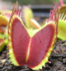
Baby VFT cilia, 40x
As above, 100x
Same VFT, cells inside the trap - plenty of chloroplasts visible
Same VFT, closed stomata with the surrounding guard cells visible
P. weser - chloroplasts, and cell walls visible
As above, a little bit closer
Amazingly, was able to see the nuclei in some of the cells. Didn't think I would be able to without some prep to the slide, but there it is! Picture isn't the greatest unfortunately.
edit- after seeing the pic on here, I think its just something else in the interstitial space, isn't within the cell so not the nuclei!
Hope you enjoyed!

Nepenthes seed closeup 40x

D. burmanii 100x

P. weser - sticky hair/glands 40x

As above 100x

Once more, 1000x

Baby VFT cilia, 40x

As above, 100x

Same VFT, cells inside the trap - plenty of chloroplasts visible

Same VFT, closed stomata with the surrounding guard cells visible
P. weser - chloroplasts, and cell walls visible

As above, a little bit closer

Amazingly, was able to see the nuclei in some of the cells. Didn't think I would be able to without some prep to the slide, but there it is! Picture isn't the greatest unfortunately.
edit- after seeing the pic on here, I think its just something else in the interstitial space, isn't within the cell so not the nuclei!
Hope you enjoyed!
Last edited by renesis on Sun Dec 27, 2009 4:57 am, edited 1 time in total.
renesis liked this
| Growlist |


 - By DragonsEye
 - By ChefDean